Electron Tomography
TEM tomography of dislocations
To model plastic deformation by dislocation dynamics, it is necessary to know the geometry of slip, i.e. the glide, climb and cross-slip planes, amongst others. A good knowledge of the geometry of dislocations interactions is also required. Electron tomography can be applied to dislocations and gives access to the real 3D structure of a dislocation microstructure. It is possible to reconstruct a small size objet (from several mm to several nm) in 3D using TEM. TEM gives projected images for different projected angles. These series of images are called « tilted series ». The angular range could reach +/- 60° for high gaps TEM polar pieces and +/- 80° for sample-holders dedicated to tomography. The more the angular range increases, the more the missing wedge decreases, and the more the reconstruction volume quality rises. Several reconstruction algorithms exist, the most common being weighted back projection (WBP) and the simultaneous iterative reconstruction technique (SIRT). They are able to transform a tilted series to a stack. The crystallographic plane distortions due to dislocations are the source of dislocation contrasts observed on TEM micrographs.
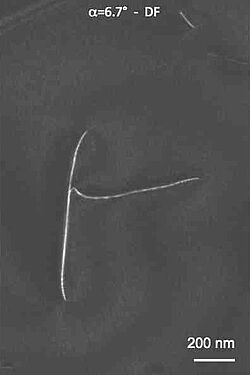
Dislocation contrasts are highly linked to the crystal orientation. Consequently, the specimen needs to be perfectly oriented with the diffraction vector aligned along the sample-holder tilt axis in order to avoid all Bragg angle variations during the tilted series acquisition (a precision of the order of a tenth of a degree is necessary). Furthermore, the micrographs have to be homogeneous in contrast to get the best 3D reconstruction quality. We perform acquisitions in the TEM weak-beam mode that gives a high signal over noise ratio. The association of a slight electron beam precession ensures to get homogeneous contrast. To increase the dislocation contrast, after acquisition it is better to filter the tilted series images using for example Kernel filtering and polynomial fit (Mussi et al., 2014). In the images here we show a weak beam dark field image of a dislocation on the right and on the left an electron tomographic reconstruction volume showing glide planes and a sessile loop lying on a climb plane.
Alexandre Mussi (Plasticité-UMET), Patrick Cordier (Plasticité-UMET)
HAADF-STEM tomography of a natural mudrock for improved fluid transport prediction
This study follows a number of publications (Song et al., 2015, Song et al., 2016, Song et al., 2016) on the multiscale imaging of a natural mudrock, where the pore network spans over several spatial scales. Former research has shown that whereas the relative pore volume of the mudrock is on average 18%, only about 2% is present down to tens of nm pore sizes. The majority of the mudrock pores is present below 10nm i.e. at a nanometric scale. Former research has allowed partial characterization of these pores, using 2D TEM and a numerical 3D image reconstruction algorithm.
This particular research aims at providing a 3D characterization of the nanometric pore network of the natural mudrock, using 3D STEM tomography. The aim is to fit the numerical 3D images of the nanometric pore network, and provide a more reliable prediction of fluid transport through the mudrock, by scale change techniques, as in [Davy and Adler, Phys Rev E, 96, 063116, 2017]. This macroscopic property needs to be fully characterized and understood, in order to analyze the radionuclide retention mechanisms. The latter are critical properties for long term radioactive waste storage.
Catherine Davy (CIMEND-UCCS), Alexandre Mussi (Plasticité-UMET), Maya Marinova (PMEL), Pierre M. Adler (UMR 7619 Metis, Sorbonne University)