
Natural Samples and Analogs
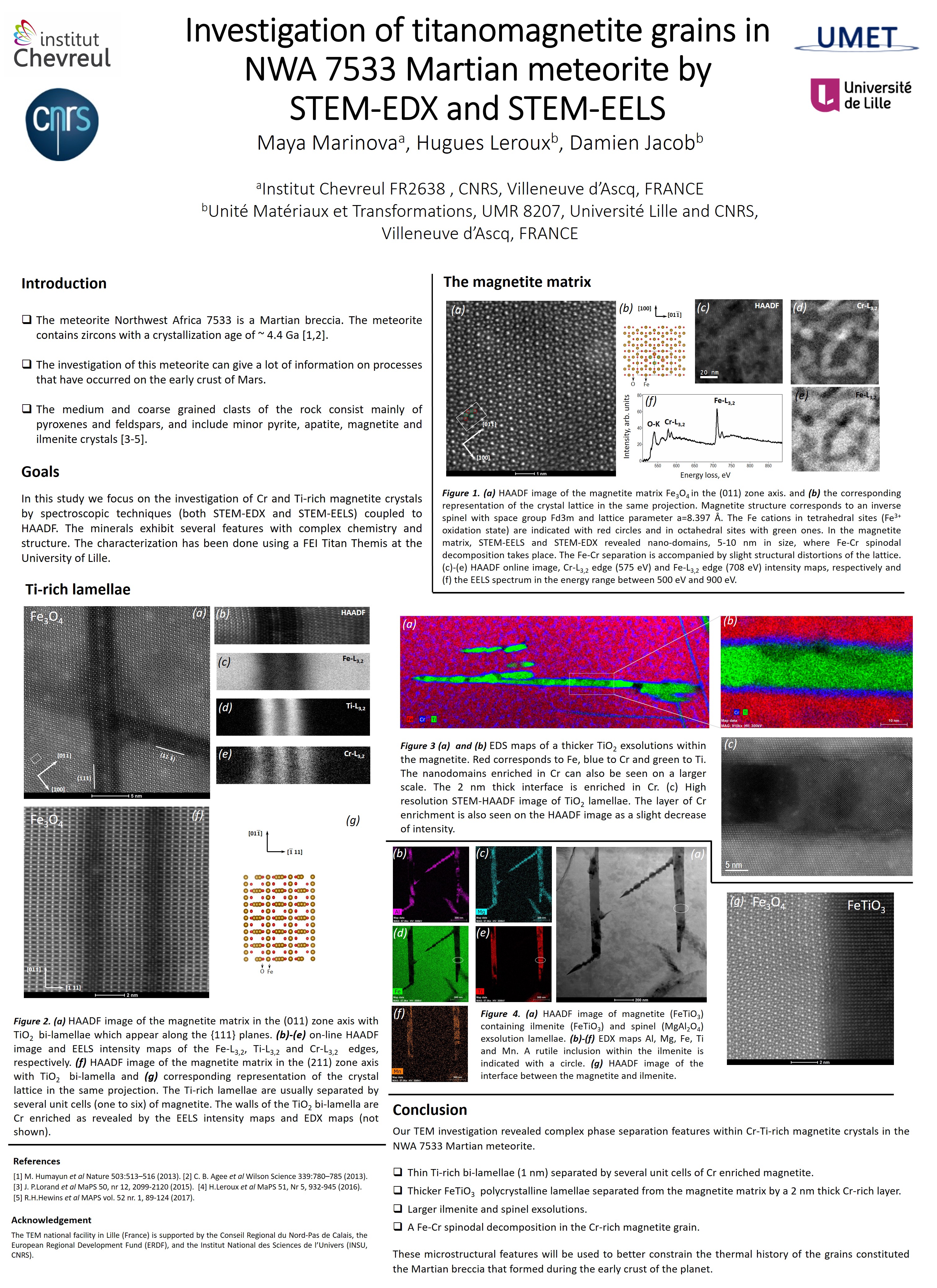
Complex phase transformation features in a Martian meteorite
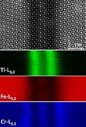
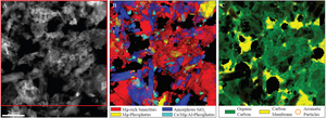
High-resolution HAADF image and EELS chemical maps from a Ti rich bilamellae in magnetite crystals in Northwest Africa 7533 Martian meteorite. The meteorite NWA 7533 has Martian origin. It is a breccia and contains zircons with a crystallization age of ~ 4.4 Ga. Unlike other Martian meteorites, this rock contains information about geological processes relative to the surface of the planet. In order to understand the thermal history and the formation of the rock, we have studied by spectroscopic techniques (EDX and EELS) coupled to high resolution HAADF and TEM images, the complex phase transformation features that appear in Ti-rich magnetite crystals. Those features include spinodal decomposition with the magnetite matrix and various exsolution slabs. The exsolutions follow {111} planes in magnetite, but display various morphology, structure and chemistry (M. Marinova et al.).
Hugues Leroux (MTP-UMET), Maya Marinova (PMEL), Damien Jacob (MTP-UMET)
Nano-phase identification in primitive chondrites by EDX
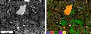
Primitive chondrites are extraterrestrial objects which present a great interest for understanding the formation of the solar system. They have been formed very early in the history of the solar system and their components have not, or slightly, changed before they have been recovered on Earth. Nevertheless, these samples are complex; they consist of an assemblage of nano-phases whose abundance can vary strongly over a few microns. TEM, and associated spectroscopies, is essential to study these objects. In order to be as representative as possible and to quantify these phase abundance variations, large-scale chemical mappings with good spatial resolution to sample nano-phases are required. The new 4-quadrant SDD detector of the TITAN THEMIS 300 makes possible to acquire this type of map with a large signal / noise ratio. This type of mapping allows to constrain processes that formed and transformed these rocks (Zanetta et al., 2019).
Pierre-Marie Zanetta (MTP-UMET), Hugues Leroux (MTP-UMET), Corentin le Guillou (MTP-UMET)
Characterization of the fundamental plastic deformation mechanisms of an olivine single crystal deformed at the lithosphere/asthenosphere boundary using electron tomography
Despite more than 50 years of research on the plastic deformation mechanisms of olivine, main constituent of the upper mantle, these mechanisms remain misunderstood. We have conducted a fundamental study of the plastic deformation of an olivine single crystal at the lithosphere/asthenosphere boundary (brittle to ductile transition of the earth mantle), near 1100°C. Olivine deforms by the glide of [100] and [001] dislocation in this temperature condition. The resulting dislocation microstructures are complex (numerous interactions and 3D dislocation configurations). We are not able to decipher these microstructures with conventional microscopy techniques; consequently we have developed dislocation electron tomography in order to see them in a new light. The video shows dislocation [100] and [001], colored in blue and orange respectively, and makes it possible to reveal the [100](001) and [001]{110} slip systems, colored in black and white respectively. Tomography enables us to identify the double cross slip mechanism and to assign a weighting for each plastic deformation mechanism, that is to say, glide, climb and cross slip. This statistic study has notably revealed the {211}, {111} and (001) preferential climb planes.
Alexander Mussi (Plasticité-UMET), Patrick Cordier (Plasticité-UMET)
Mapping organics association with phyllosilicates by advanced EDX data processing
Understanding the physics and chemistry behind the nanometer-scale association of organics and hydrated silicates (phyllosilicate) is of the highest importance in various fields such as soils, sediments and meteorites sciences. To reveal the nature of the interaction between them, we developed a protocol to “cryo-microtome” powder samples and map them with the silicon drift, windowless EDS detectors. The latter have a high sensitivity at the carbon K-edge energy which allows efficient mapping of carbon simultaneously with other elements. However, emitted carbon X-rays can be strongly re-absorbed within the sample and careful absorption correction is crucial. In order to correct for these absorption issues, we process the EDS hyperspectral data (HyperSpy), including pixel per pixel absorption correction, elemental quantification and creation of phase maps. Our results highlight the association of organics with specific phyllosilicates. In the figure we show experimental product of a smectite-RNA mixture heated at 200°C for 7 days in a CO2 atmosphere (Viennet et al., submitted). The STEM-HAADF image shows the porosity and the scale of the individual particles. The phase map reveals the presence of secondary phases, while the organics map reveals that they are preferentially associated to smectite. The color intensity is a function of the phase abundance at each pixel (organics included) (Viennet et al., 2019, Jacquemot et al., 2019, Viennet et al., 2020).
Corentin Le Guillou (MTP-UMET), in collaboration with Jean-Christophe Viennet, Sylvain Bernard, Pierre Jacquemot and Maguy Jaber (MNHN et Sorbonne Université, Paris, France)
EDS quantification of the altered layer composition of a glass designed for nuclear waste storage
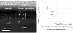
Borosilicate glasses are host candidates for the underground storage of nuclear waste. However, they will be submitted to interaction with water in the storage galleries, where uclides could be released in the environments during alteration. Therefore the kinetics of the glass alteration must be carefully studied. Here, we analyzed and quantified a glass altered in vapor conditions (90°C, 95% relative humidity). The alteration layers are extremely thin and their stability under the beam is weak. The composition profile of these complex multi-layered material could nevertheless be obtained. In the figure STEM HAADF image of a borosilicate glass altered at 90°C in vapor conditions, and associated composition profile obtained by quantification of EDS hyperspectral maps (Narayanasamy et al., 2019).
Corentin Le Guillou (MTP-UMET), in collaboration with Sathya Narayanasamy, P. Jollivet, N. Godon, F. Angeli, S. Gin, M. Cabié, J. Cambedouzou, A. Abdelouas (CEA, Marcoule, France)
Characterization of the deformation mechanisms of the earth low mantle phases using orientation mapping
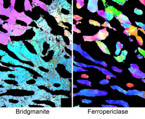
A large specimen (a fraction of mm3) from a mixture of bridgmanite and ferropericlase, main constituent of the earth lower mantle, has been highly deformed by torsion for the first time under lower mantle pressure and temperature conditions. This experiment, conducted at the Geology and geophysics laboratory of Yale University, is a real technical challenge. Consequently, the TEM (transmission electron microscopy) analysis of deformation microstructures of this exceptional sample is paramount. However, we have confronted two obstacles. On the one hand, the electron beam extreme sensibility of bridgmanite, and on the other hand, the high dislocation density of ferropericlase making the characterization of individual dislocation impossible. Thanks to fast electron beam scanning with micro-probe mode, the ASTAR technique makes it possible to preserve the structures of sensitive phases to electron beam. It also enables us to obtain orientation maps and so misorientation maps. Yet misorientations give access to deformations since dislocations generate misorientations. In doing so, we have associated EBSD (Electron BackScatter Diffraction) tools, that is to say GOS (Grain Orientation Spread), GROD (Grain Reference Orientation Deviation) and KAM (Kernel Average Misorientation), to the orientation maps obtained by TEM. High deformation heterogeneity and grain fragmentations of ferropericlase have been characterized by GOS and GROD mapping. Furthermore, ferropericlase KAM distributions have shown a lack of recrystallization despite strains of hundreds of percent. KAM mappings of bridgmanite have revealed the occurrence of misorientation concentrated zones corresponding to amorphous lamellae. Bridgmanite seems to deform by amorphous lamellae formation where shear is concentrated, and not by dislocation motion. ASTAR technique associated with tools employed by the EBSD community users enables us to get these original results. (Nzogang et al., 2018).
Alexander Mussi (Plasticité-UMET), Patrick Cordier (Plasticité-UMET), Billy Nzogang (Plasticité-UMET)